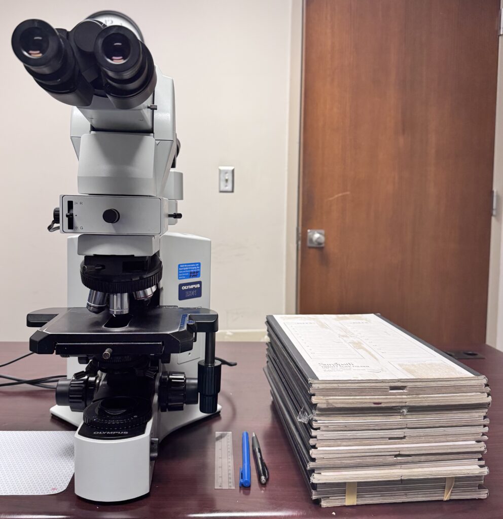
When you arrive in the morning to a huge stack of slide folders, it’s very tempting to want to jump right into reviewing slides to get through them as efficiently as possible. But, efficiency isn’t the only goal or even the primary goal. The primary goal is to get the right diagnosis the first time, while also being efficient. A systematic approach to reviewing cases is a helpful strategy for accurate and efficient sign out. Here is an approach that works for me based on some best practices that I’ve learned over the years:
1. Verify the patient demographics
Verify that the case number matches the patient demographics. Block/slide mixups do occur and it’s much easier to resolve these errors prior to signing out a case.
2. Perform block inventory
Each case will consist of one or multiple parts and each part will consist of one or more slides. It’s not uncommon for parts of the same case to be received, grossed or processed at different times (e.g. parts for frozen section vs. permanent, decalcified specimens).
Verify that you have received all parts and slides for the case.
3. Review the clinical history
Ask yourself: What is the clinical question?
Then consider, what information would help me to answer this clinical question? For example, if the case is a liver biopsy – the clinical question could be “Neoplasm? Benign or malignant?” or “Cirrhosis? What stage?” If the question is one about a mass lesion, then reviewing recent clinic notes for relevant cancer history and radiology reports is important. If the question is one of medical liver disease, then results from chemistry labs and serologies is important.
Perform a thorough, but not comprehensive, review of the clinical history including any previous pathology reports. In the world of clinicians, this is similar to interviewing the patient for past medical history, social history, and family history. You don’t need to read it all but you do need to know what’s going on with the patient – consider it the “rounds” of pathology.
4. Read the gross description and summary of sections
Most pathologists are delighted when they no longer need to gross specimens (some pathologists still have the privilege) but the gross description is still important.
Review the gross description to understand the findings. If you’re a visual learner, it can be helpful to sketch the findings on scrap paper. I find this particularly helpful with skin and breast resections when the assessment and measurement of margins can be complicated. The summary of sections is important to not only know what exactly each slide represents but also to know that the appropriate sections were taken (e.g. bowel resections should include proximal, distal, and radial/mesenteric margins).
5. Review the slides
6. Formulate a differential diagnosis
Many cases are very straightforward but, if it’s not a slam dunk, picture-matching diagnosis, then it’s prudent to always consider a differential diagnosis. Here are some questions to ask yourself:
What do I think this is?
What do the clinicians think this is?
What else could it be?
What diagnosis, if missed, could be catastrophic?
For example, I recently had a case of a lung biopsy of a solitary lung nodule. It had most of the classic features of primary lung adenocarcinoma but looked just a little unusual. It would have been very easy to sign it out as “adenocarcinoma” and move on to the next case but I paused to consider if this could be something else. The immediate clinical history was unrevealing but I dug deeper into the medical record and discovered that the patient had a remote history of endometrial adenocarcinoma. The reasonable approach to a case like this is to order immunohistochemical stains (see next step) but this requires you to pause to consider that next step. The final diagnosis was metastatic endometrioid endometrial adenocarcinoma.
7. Order immunohistochemical (IHC) stains, if needed
Not every case requires an immunohistochemical stain. Avoid being the pathologist who relies too much on stains as they can lead you astray. It’s no different than a clinician who indiscriminately orders clinical lab tests without thought and then has to interpret data that only presents noise rather than signal. H&E is your best friend.
This being said, IHC stains are very helpful in the practice of pathology and, fortunately, the panel of available stains continues to increase. When ordering stains, consider stains to help confirm the diagnosis as well as exclude other diagnoses in your differential diagnosis.
8. Finalize the diagnosis
Is the final diagnosis clear and concise (i.e.. Will the clinician understand without a clarifying phone call)?
Did you comment on any additional studies performed?
Did you document any intra- or extradepartmental consultations?
Did you include any pertinent positives or negatives (e.g. in a breast biopsy to assess calcifications, comment on the presence or absence for radiologic concordance)?
9. Proofread!
Always, always, always review your diagnosis for typos. This is critically important whether you’re typing, dictating, transcribing, or using macros/SmartText.