History: 44 y/o female presents with biliary colic and undergoes a cholecystectomy
Histology:
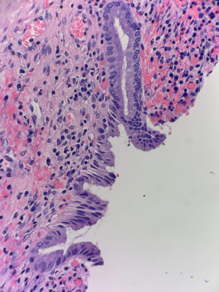
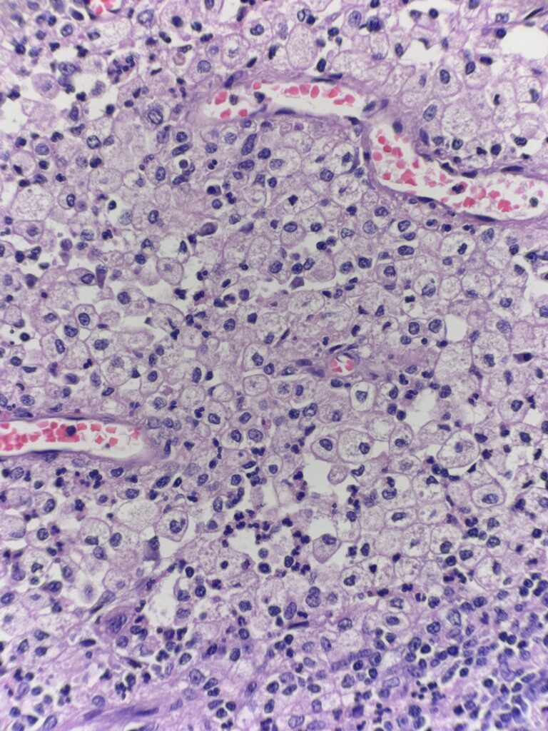
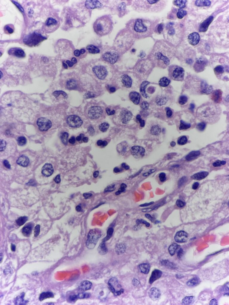
Diagnosis: Xanthogranulomatous cholecystitis
The first image shows gallbladder mucosa with chronic inflammation and reactive epithelial changes. The following images show regions deeper in the submucosa and muscularis consisting of nodular aggregates of foamy histiocytes. These cells are positive for CD68 and negative for pankeratin – to differentiate this inflammatory entity from an infiltrating carcinoma.