Clinical history: 75 year-old female with intestinal metaplasia seen on previous random gastric biopsy
Site: Stomach (body)
Histology:
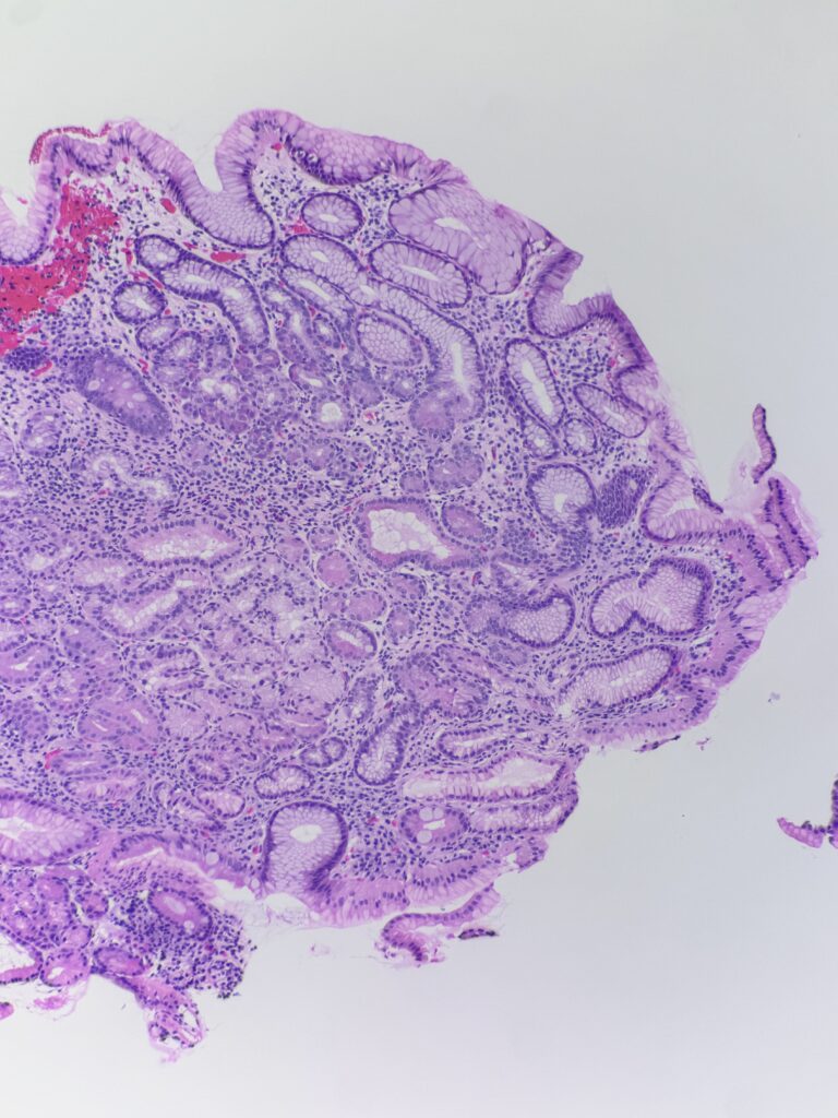
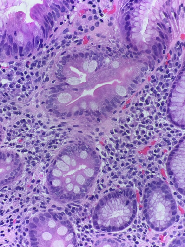
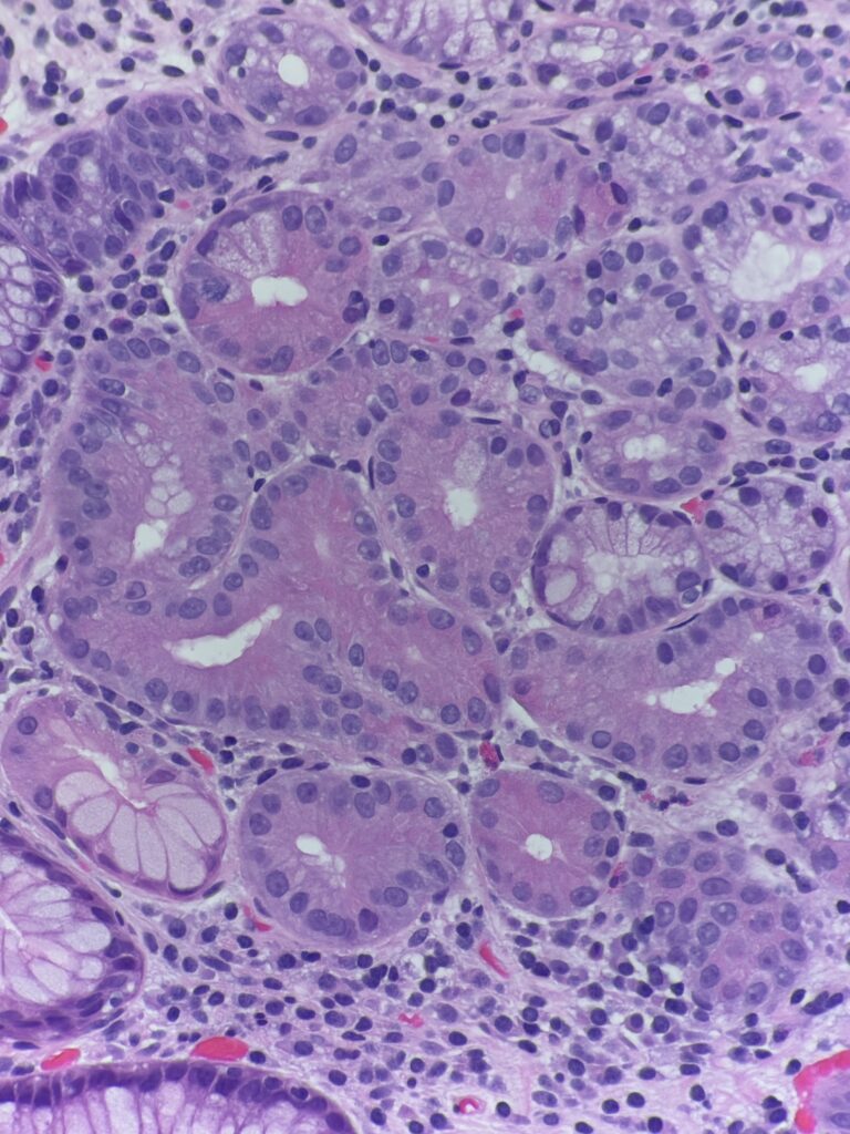
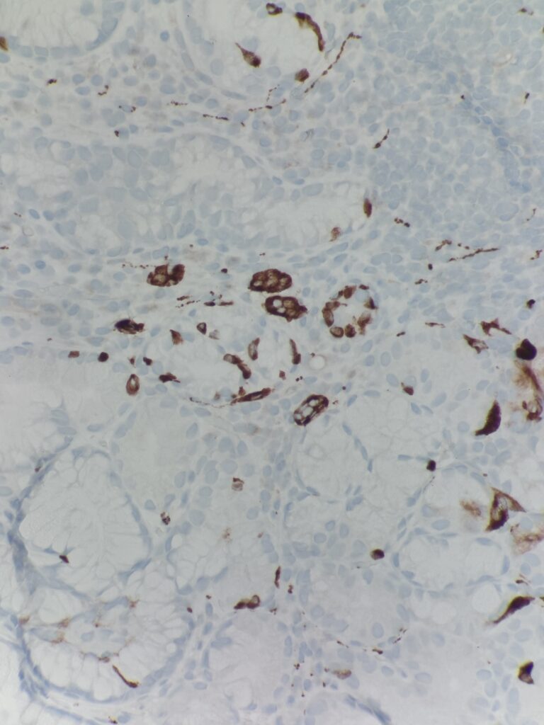
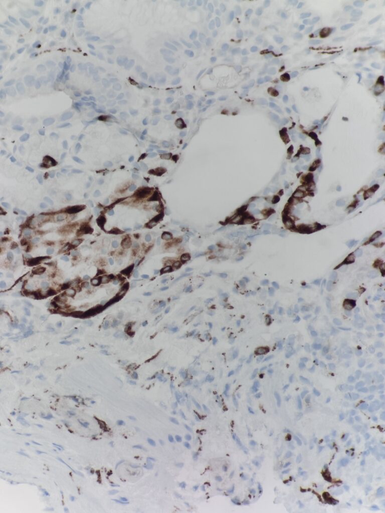
Diagnosis: Autoimmune metaplastic atrophic gastritis
Diagnostic notes: As compared to H. pylori gastritis, the inflammation is predominantly in base of the mucosa extending to the superficial mucosa. There is loss of oxyntic glands, intestinal metaplasia, pancreatic acinar metaplasia and nodular and linear enterochromaffin like (ECL) cell hyperplasia. Gastrin stain can be used to differentiate antral vs. body mucosa – gastrin stain is positive in the antrum.
- Final report notes:
- +/- Helicobacter organisms
- +/- Intestinal metaplasia (if yes, +/- dysplasia)
- Clinical correlation: recommend serologic testing for anti-parietal cell and anti-intrinsic factor antibodies